Article

Influence of COMT (rs4680) and DRD2 (rs1076560, rs1800497) Gene Polymorphisms on Safety and Efficacy of Methylphenidate Treatment in Children with Fetal Alcohol Spectrum Disorders

by
Małgorzata Śmiarowska
1,
Bogusław Brzuchalski
1,
Elżbieta Grzywacz
2,
Damian Malinowski
1,
Anna Machoy-Mokrzyńska
2,
Anna Pierzchlińska
1 and
Monika Białecka
1,*
1
Department of Pharmacokinetics and Therapeutic Drug Monitoring, Pomeranian Medical University, Aleja Powstancόw Wielkopolskich 72 St., 70-111 Szczecin, Poland
2
Department of Experimental and Clinical Pharmacology, Pomeranian Medical University, Aleja Powstancόw Wielkopolskich 72 St., 70-111 Szczecin, Poland
*
Author to whom correspondence should be addressed.
Int. J. Environ. Res. Public Health 2022, 19(8), 4479; https://doi.org/10.3390/ijerph19084479
Submission received: 7 February 2022 / Revised: 29 March 2022 / Accepted: 6 April 2022 / Published: 8 April 2022
(This article belongs to the Special Issue Pregnancy and Child Health)

Abstract

:
Fetal alcohol spectrum disorders (FASD) in a course of high prenatal alcohol exposure (hPAE) are among the most common causes of developmental disorders. The main reason for pharmacological treatment of FASD children is attention deficit hyperactivity disorder (ADHD), and methylphenidate (MPH) is the drug of choice. The aim of the study was to assess whether children born of hPAE with ADHD, with or without morphological FASD, differ in terms of catechol-O-methyltransferase (COMT) and dopamine receptor D2 (DRD2) gene polymorphisms, and if genetic predisposition affects response and safety of MPH treatment. The polymorphisms of COMT (rs4680) and DRD2 (rs1076560, rs1800497) were analyzed in DNA samples. A borderline significance was found for the correlation between MPH side effects and the G allele of COMT (rs4680) (p = 0.04994) in all ADHD children. No effect of COMT (rs4680) and DRD2 (rs1076560, rs1800497) polymorphisms and the treatment efficacy was observed. The analyzed DRD2 and COMT gene polymorphisms seem to play no role in MPH efficacy in ADHD children with hPAE, while low-activity COMT (Met158) variant carriers may be more intolerant to MPH. The MPH treatment is effective in ADHD independent of FASD, although the ADHD-FASD variant requires higher doses to be successful. These results may help in optimization and individualization in child psychiatry.

1. Introduction

Alcohol consumption during pregnancy may lead to permanent damage in the offspring, resulting in, for example, Fetal Alcohol Spectrum Disorders (FASD). FASD include Fetal Alcohol Syndrome (FAS), partial Fetal Alcohol Syndrome (pFAS), alcohol-related neurodevelopmental disorders (ARND), and alcohol-related birth defects (ARBD) [1]. FAS is believed to be the most common teratogenically induced, nonhereditary form of mental deficiency throughout the Western world [2,3,4]. It is associated with reduced intelligence, attention disorders, neuropsychological deficits, physical abnormalities including facial dysmorphology, sleep disorders, and behavioral problems [5,6,7].
According to worldwide studies, alcohol consumption among women of childbearing age is increasing. At the same time, a large proportion of pregnancies are unplanned, which leads to more frequent cases of prenatal alcohol exposure (PAE) which may be explained by harmful and sustained alcohol intake during any trimester of pregnancy which, in consequence, may produce morphological, biochemical, and epigenetic (among others) consequences in the fetus. Alcohol has a prolonged effect in pregnancy in terms of abundance and efficiency caused by the differences in maternal and fetal enzymes, oxidative stress/reactive oxidative species (ROS) and, additionally, reduced elimination. In 120 min after the mother’s alcohol intoxication, its concentration in her blood increases to maximum (blood alcohol content BAC). Owing to placenta transport of alcohol, its concentration after 120 min gets maximal BAC which is nearly the same as maternal. During pregnancy there is no safe dose of alcohol and most of PAE state in the first trimester of pregnancy [8]. The structural and neurobehavioral consequences associated with PAE are the most teratogenic in the brain. Post-mortem and brain imaging studies showed reductions and anomalies in the brain size and shape (seen as microcephaly and hydrocephalus), especially in the cerebellum, basal ganglia, and corpus callosum, as well as grey matter heterotopia and neurochemical abnormalities [9,10].
The multiple clinical consequences of the prefrontal cortex area damage in FASD children involve cognitive domains (general intelligence, visual perception and construction and learning and memory), motor functions and activity levels, language skills and adaptive functioning [11,12,13,14]. In addition, PAE has a high comorbidity rate with other learning and behavioral disorders [15]. The attention deficit hyperactivity disorder (ADHD) is one of them [16,17,18,19]. A correlation between FASD and ADHD has become a focus of special scientific attention. It was established that FASD constitutes one of the main susceptibility factors for developing ADHD (relative risk 7.6; attributable risk 86.8%). Similarly, displaying the symptoms of ADHD increases the probability of diagnosing FASD (relative risk 13.28; attributable risk 92.5%) [20]. There are numerous, likely interacting, genetic and environmental factors involved in mediating ADHD and its severity [21,22,23]. Since FASD and ADHD are characterized by similar phenotype expression, prevalence, and detrimental impact they have on the child’s development, a better understanding of neurobiological underpinnings of PAE-related ADHD may lead to a faster diagnosis and more effective pharmacological treatment of ADHD symptoms [24,25].
In addition to psychological and behavioral therapies, NICE guidelines (National Institute for Health and Care Excellence) allow a pharmacological treatment of individuals older than 5 years with a severe presentation of ADHD. Stimulants, such as methylphenidate (MPH), are considered to be the most effective medications for the improvement in ADHD symptoms and MPH is often the first-choice substance owing to its effectiveness and safety, as demonstrated in several studies [26,27].
The mechanism by means of which MPH reduces symptoms in ADHD is not completely clear. One of the most known conceptions of the ADHD pathogenesis is the one related to delayed dopaminergic system maturation in the brain, and medication such as MPH can improve DA transmission. It seems to be similar to cocaine and involved in the central stimulation of catecholamines. It is believed that MPH binds to dopamine transporter (DAT) and norepinephrine transporter (NET) in the presynaptic membrane. In this way, it blocks the reuptake of dopamine and norepinephrine and in turn increases intrasynaptic concentrations of DA dopamine and norepinephrine (NE) and increases them in the synaptic cleft [28]. Depending on the brain region activation, these catecholamines (especially DA) have a specific clinical manifestation; for example, in the prefrontal cortex (PFC) they are more responsible for cognitive control–executive functions (inattentive type of patients), while in the subcortical brain regions (a striatal–frontal loop) they are more strongly associated with motivation and reward (the hyperactive and combined types) [28]. The other mechanism by means of which MPH affects the hyperactivity and emotional disturbances (aggression and anxiety) in ADHD is probably connected with its indirect stimulation of the dopamine D2 postsynaptic receptor (DR2) [29,30,31]. PFC contributes to the unusual property of the DA system, and there is a hypothesis of exceptional PFC’s vulnerability to environmental and genetic variation [32]. As mentioned before, not only environmental but also genetic factors may be involved in ADHD etiology and its severity. Many studies have extensively investigated the role of polymorphisms in the DR2 gene (DRD2), e.g., Taq1A (rs1800497), C957T (rs6277), and −141Cins/del (rs1799732), in neuropsychiatric disorders such as schizophrenia, alcoholism, Parkinson’s disease, substance addiction, and ADHD [33,34,35,36,37,38]. A meta-analysis conducted by Wu et al. identified a significant association between ADHD and Taq1A polymorphism of DRD2 (OR 1.65, 95% CI 1.05–2.58, p < 0.0001) [39]. The result was supported by a subsequent meta-analysis (95% CI = 1.068–2.984, p = 0.027) [38]. Interestingly, the authors observed differences between ethnicities: the polymorphism was associated with ADHD in Africans, but not in East Asians or Caucasians. Aggressive behavior, which is very close to impulsiveness, is one of the clinical symptoms met in ADHD. The study conducted by Zai at al. explored the role of dopaminergic system genes in the etiology of aggressive behavior in children [40]. The authors discovered a significant association between A-241G (rs1799978), rs1079598, and Taq1A (rs1800497) of DRD2 polymorphisms and childhood aggression. Genetic variation in enzymes that act on the biosynthesis or degradation of DA and its metabolites may influence the effectiveness of MPH in ADHD treatment [41]. Catechol-O-methyltransferase (COMT) inactivates biologically active or toxic catecholamines such as DA and norepinephrine (NE) and plays a role in the metabolism of drugs (L-dopa). Differences in COMT activity and genotype may determine individual variations in the therapeutic response to drugs, whose mechanism of action are linked to DA and NE levels. A single COMT gene, located on chromosome 22q11, encodes both the acid-soluble (S-COMT) and membrane-bound (MB-COMT) forms of this enzyme. The most studied polymorphism of the COMT gene is a G to A transition at codon 158 [Val158Met; rs4680]. The transition resulting in the substitution of methionine for valine has been linked to low COMT enzyme activity and is designated as the L (low-activity) allele, in contrast to the H (high-activity) allele [42].
Jung et al. found a correlation between the COMT Val158Met polymorphism and cortical thickness and surface area in children with ADHD, cortico-cerebellar executive function network, or stimulants’ efficacy [43]. The impact of genetic factors on ADHD pathogenesis, as well as on the efficacy and safety of MPH treatment in ADHD patients, has been analyzed, however, without any conclusive results [44,45,46,47]. The study aimed at assessing whether children with ADHD, with or without FASD, differ in terms of catechol-O-methyltransferase (COMT, rs4680) and dopamine receptor D2 (DRD2, rs1076560 rs1800497) gene polymorphisms, and if the genetic predisposition affects the response and safety of the methylphenidate (MPH) treatment in children with ADHD and high prenatal alcohol exposure (hPAE) with or without morphological features FASD, according to the Washington Questionnaire.

2. Materials and Methods
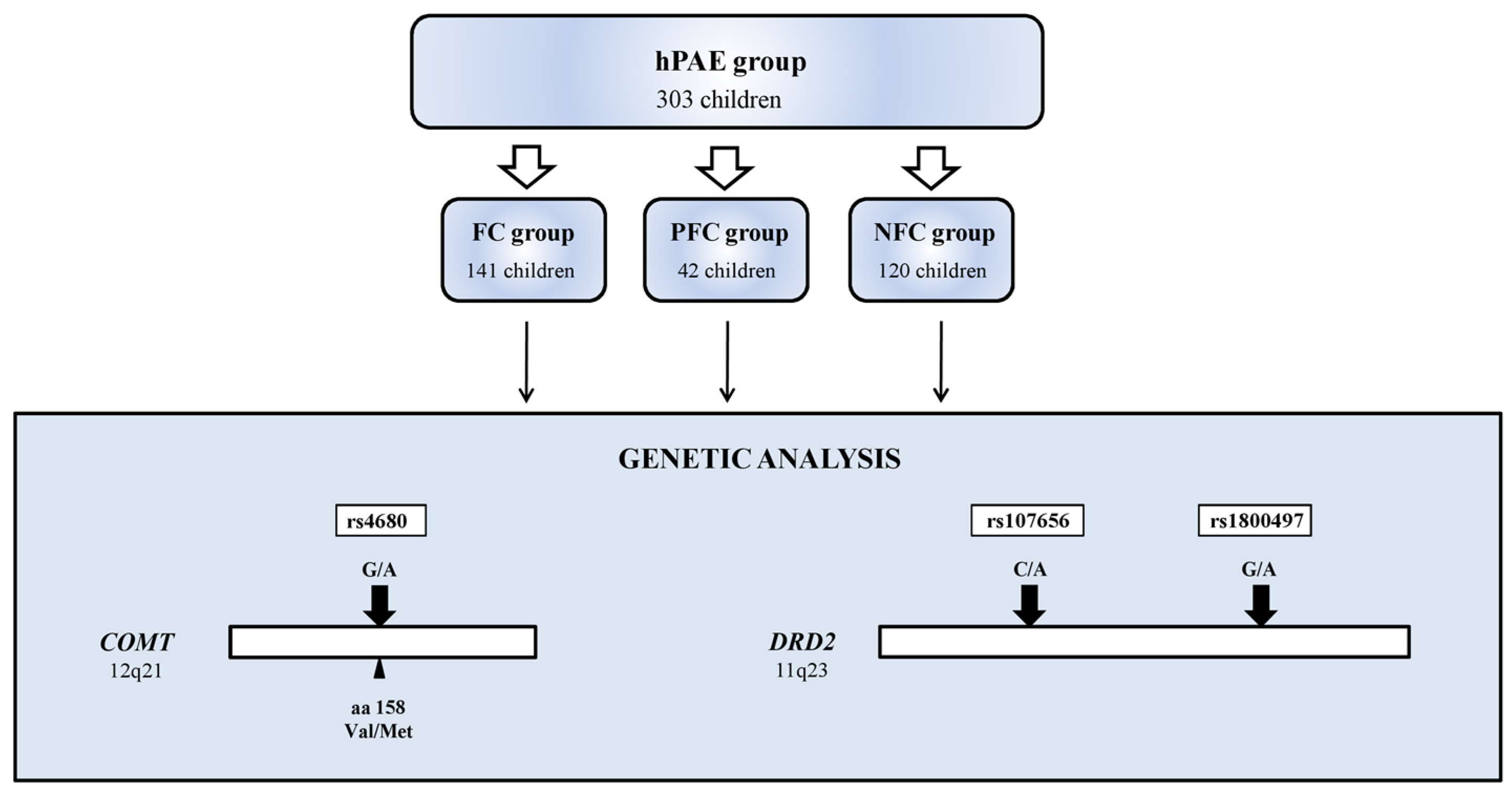
2.1. Study Group

Three hundred and three children were included in the study: 213 girls (mean age 8.59 ± 4.77 years) and 90 boys (mean age 9.77 ± 5.14 years). The study group consisted of 303 hPAE children, with 183 ones diagnosed with FASD (partial FAS children—PFC, n = 42, and FAS children—FC, n = 141) and NFC children without the presence of evident morphological FAS changes unless positive history of hPAE (non-morphological FAS children—NFC, n = 120) (Table 1, Figure 1). All children enrolled in the study met the criteria for the occurrence of high alcohol exposure during fetal life/(hPAE). PAE was confirmed by the mother’s medical history or by objectively documented background survey (e.g., mother’s stay in a sobering station or in a detoxification ward during pregnancy). The clinical diagnosis of FASD was based on a 4-digit diagnostic questionnaire according to a validated validation of the Polish version of the Washington Questionnaire for the Assessment of Fetal Alcohol Spectrum Disorders [48]. It is worth pointing out that all of FASD children enrolled to the present study had severe OUN lesions or disturbed functionality which were not related to genetic or environmental factors (both FC and PFC received 3 or 4 points on the 4th degree scale) and they were short-statured or had specific face anomalies. They are generally moderate (3rd and 2nd degree) ARND according to the Washington Questionnaire.
In the total of 303 hPAE children enrolled for the study, 114 children met the criteria for ADHD diagnosis according to the American Psychiatric Association (APA) DSM-IV criteria [49] and the ICD-10 classification of Mental and Behavioral Disorders: Clinical Description and Diagnostic Guidelines [50]. The quantitative assessment of the severity of symptoms was performed according to the questionnaire developed by Wolańczyk and Kołakowski based on the DSM-IV rating scale (RS) and ICD-10 criteria which is not an international diagnostic valid instrument for ADHD screening, but a very approachable and popular form of interviewing and simple in its interpretation form in common practice (the pattern is available in Supplementary Materials) [51]. It examines three areas of the child’s dysfunction using separate scales: (I). attention deficit includes nine descriptions of problem behaviors, (II). hyperactivity—five, (III). impulsivity—four. The severity of each symptom is measured from 0 to 3 points (0—the symptom occurs never or very rarely, 1—sometimes, 2—often, 3—very often). Therefore, in each of the scales of the examined area, the patient may obtain a maximum of 27 in I scale, 15 in II scale, and 12 points in III scale, respectively. The numerical values served as a reference for the assessment of the intensity of attention deficit, hyperactivity, impulsivity before (BT) and after the treatment (AT) of MPH.
According to the DSM-IV RS criteria, the examined children were divided into 3 subgroups (subtypes) depending on the predominance of the clinical picture (attention deficit disorder vs. activity/impulsivity assessed together): attention deficit/hyperactivity disorder (ADHD-A), hyperactivity/hyperimpulsivity disorder (ADHD-HI), and a mixed form (ADHD-M) when criteria of both subtypes are balanced.
The ICD-10 classification was used to investigate the contribution of the child’s environmental functioning conditions to the clinical manifestation of hyperkinetic syndrome (F90.0/ADHD) symptoms to consider all probable factors playing a role in its pathogenesis, except biological pathways of catecholamines involved in it. According to the diagnosis criteria, F90.0 child’s impaired functioning includes at least 2 out of 3 areas examined, including peer group, school, and home, which are observed at the same time by minimum 2 out of 3 assessments from the perspective of various people from the child’s environment.
The criteria for the patient’s exclusion from the study were psychiatric disorders: psychotic (present and in medical history), significant mood and anxiety disorders requiring pharmacological treatment, psychoactive substance (SPA) addiction, risky and harmful use of SPA (present and in medical history); and developmental disorders: autism spectrum, profound and significant mental retardation, genetic diseases, severe and uncompensated somatic diseases (endocrinological, cardiovascular, renal, neoplastic, autoimmune, anorexia), and organic diseases with clinical manifestation (epilepsy). A slight shortage of weight and height according to age, but within normal limits (>10th centile), and a history of depressive episodes and emotional and behavioral disorders did not exclude patients from the study.

2.2. Eligibility for MPH Treatment

Within the study group, i.e., 303 children with hPAE, 114 children were included in the methylphenidate treatment. The children were older than 6 years old, with a diagnosis of ADHD, who did not benefit from cognitive behavioral therapy (CBT) for last 6 months. The 6 months psychotherapy condition did not apply to children whose ADHD symptoms were severe and caused dysfunctional environmental functioning. Before the introduction of the drug, each patient underwent examinations: electrocardiography, blood pressure, heart rate, weight, and height. Initial doses of 5 mg and 10 mg MPH in short-acting forms were used to assess drug tolerability (severity of nausea, abdominal pain, decreased appetite, and cardiovascular effects) and appropriateness of further administration. Established MPH treatment was continued with prolonged-release forms approved for use in Poland: Medikinet 10 CR (10 mg methylphenidate hydrochloride, by Medice) and Concerta 18 (18 mg methylphenidate, by Janssen). The doses were increased twofold: Medikinet 20 CR and Concerta 36 mg, which were the therapeutic doses and the basis for analysis in the current study. The maximum dose (Medikinet 60 CR and Concerta 54 mg) was used only in individual cases, which did not affect the general observation. The choice between Medikinet CR vs. Concerta was based on the patient’s daily requirement for adjustments and according to the drug’s half-life (TT1/2 = 8 h vs. TT1/2 = 12 h, respectively).

2.3. Assessment of Efficacy and Safety of MPH Treatment

The evaluation of the efficacy and tolerability of methylphenidate was carried out based on the data from the parents or guardians and other persons from the child’s functioning environment (teacher, guidance counselor, or educator in the institution). To measure the therapeutic effect of MPH, the questionnaires based on DSM-IV RS, described in the methodological section, were applied. The first assessment was performed at time “0” at the qualification for treatment. The treatment effect was assessed at three time points (after 8, 12, and 24 weeks), although the period of the first 8 weeks was the best to estimate the control exam to be sure that all children followed the established regimen. The results were effective when the number of problematic symptoms in terms of the examined scale (attention deficits, hyperactivity, and impulsivity, respectively) was reduced by more than 40% in at least two of the examined environments.
The assessment of MPH adverse effects (AEs) was based on the occurrence of symptoms such as: loss of appetite and weight loss (greater than 10% in 4 weeks), nausea, abdominal pain, headache, sleep problems, cardiovascular and emotional disorders (increased fear, anxiety, aggression and autoaggression), fatigue, and skin lesions. The criterion for discontinuation of the treatment due to AEs (according to the characteristics of the product) was the occurrence of any of the following symptoms: cardiac arrhythmia, appearance of suicidal thoughts or intentions, perceptual disturbances (visual, auditory, sensory hallucinations), attacks of uncontrolled aggression and agitation (which had not been present before the drug was started), motor and/or vocal tics, sudden increase in body temperature, or morphological changes in the blood picture (decrease in the cell count). No such severe adverse effects during MPH treatment appeared to exclude any patient from the observation.
Written consents were obtained from the legal guardians of all the studied children, and children who were 16 years of age or older. The study was approved by the local Bioethics Committee at the Pomeranian Medical University in Szczecin (the approval of the Bioethics Committee of the Pomeranian Medical University in Szczecin from 5 October 2015, No. KB-0012/95/15).

2.4. Genetic Analysis

Genomic DNA was extracted from buccal swab samples, and cells from mucus membrane were collected using a sterile synthetic swab. Prior to the isolation, swabs were stored in a preservative buffer at +4 °C. Extraction was performed using Genomic Micro AX SWAB Gravity kit (A&A Biotechnology, Gdynia, Poland) following manufacturer’s protocol. DNA was subsequently standardized to equal concentrations of 10 ng/μL, based on spectrophotometric absorbance measurement (260/280 nm). List of the SNP assays is shown in the table below (Table 2).

2.5. Genotyping

Genotyping for COMT rs4680, DRD2 rs1076560, and rs1800497 SNPs was performed using pre-validated allelic discrimination TaqMan real-time PCR assays (Life Technologies, Waltham, MA, USA), and TaqMan GTXpress Master Mix (Life Technologies, Waltham, MA, USA). All reactions were run in a final volume of 12 μL (reaction temperature profile: 95 °C, 20 s, 40 × 95 °C 1 s/60 °C, 20 s).
Fluorescence data were captured using the ViiA7 Real-Time PCR System (Applied Biosystems, Waltham, MA, USA) after 40 reaction cycles. Specific genotypes were assigned to individual samples after the analysis with TaqMan Genotyper software (Thermo Fisher Scientific, Waltham, MA, USA).

2.6. Statistical Analysis

The chi2 (χ2–Pearson’s test) and/or Fisher’s exact test were used to compare qualitative variables between genotype groups, alleles, as well as clinical data in genotype groups. For genotype and allele distribution frequencies Hardy–Weinberg equilibrium was calculated. Age distribution according to SNP genotypes was performed using Kruskal–Wallis test. p < 0.05 was considered statistically significant. Statistical analysis was performed using STATISTICA PL, ver. 13.1 software (StatSoft, Inc. 2016, Tulsa, OK, USA, STATISTICA-data analysis software system, version 13.1) software. Haplotype reconstruction and linkage disequilibrium analysis were performed by means of HaploView 4.2 software.

3. Results

In the group of 303 children with hPAE enrolled for the study, 114 were diagnosed with ADHD. Seventy-one of them had morphological symptoms of FASD (partial FAS children—PFC and FAS children—FC) and 43 children did not show any morphological changes of FASD (no FASD children—NFC). The clinical picture of children with ADHD included features of attention deficit disorder and hyperactivity with attention deficit (ADHD-A), and attention deficit disorder and hyperactivity with predominantly hyperactivity and impulsivity (ADHD-HI). No individuals were diagnosed with the mixed form of ADHD (ADHD-M).
According to the protocol, MPH was introduced in 114 children with ADHD symptoms. The mean daily dose of MPH was 27.37 mg. However, in the FASD subgroup the established doses were significantly higher compared to the NFC subgroup (n = 71, dose 31.6 mg vs. n = 43, dose 18.67 mg, respectively, p = 0.0112).
In both the FASD/ADHD and NFC/ADHD groups, pharmacological treatment was effective in >90% of the patients (n = 104 successfully treated patients). No improvement was found in three patients treated with MPH, and in seven children, adverse effects (AEs) occurred, and the experiment was discontinued.
The efficacy of MPH among FASD/ADHD children was observed in the decreased symptoms of hyperactivity and impulsivity (p < 0.0001), with no improvement in attention deficits (p = 0.2024). In contrast, NFC/ADHD children showed statistically significant improvement in attention and reduction in hyperactivity (p < 0.001 and p = 0.0163, respectively) (Table 3).
Adverse side effects occurred in 7 out of 114 patients treated with MPH. Symptoms of intolerance appeared quickly after the drug introduction and were associated with noradrenergic and dopaminergic overstimulation (transient tachycardia, loss of appetite, headache, stomach pain, insomnia trouble, anxiety, and rise in autoaggression and aggression). Their clinical presentation and severity decreased after introduction of a modified form of MPH. No cardiotoxic effects or life-threatening symptoms were reported which could force discontinuation of the therapy in this group.
No associations were noted for the analyzed gene polymorphisms. p-values for allele frequency comparison and odds ratio (OR) for different inheritance models are given in Table 4 and Table 5, while full genotyping data is provided in Supplementary Table S1. The distribution of genotypes in accordance with the Hardy–Weinberg equilibrium was observed in the case of all studied SNPs. Separate subgroup analysis within the FASD group (FAS vs. NFC, pFAS vs. NFC, FAS + pFAS vs. NFC) show no significance (data not shown).
Considering linkage disequilibria between SNPs located within the same gene, the frequencies of haplotypes reconstituted from genotyping results for two DRD2 SNPs (rs1076560 and rs1800497) were compared between the FASD and NFC groups. A-A and A-C DRD2 haplotypes were less frequent among NFC children compared to the FASD (0.154 vs. 0.183, and 0.013 vs. 0.028, not significant), while the major G-C haplotype was more frequent among NFC children (0.829 vs. 0.784, not significant). Linkage disequilibria and haplotype frequencies are provided in Supplementary Tables S2 and S3.
In children without morphological signs of FASD who carried the minor A allele of the COMT rs4680:G > A polymorphism, the clinical picture was more often associated with emotional anxiety and/or depressive disorders, and MPH was the drug of choice. The mean age in this group was 8.45 years (SD = 3.56 years) and the mean dose used was 11.34 mg/d.
There was a statistically significant association between the occurrence of adverse reactions in children treated with MPH and the presence of the COMT rs4680 minor A allele (p < 0.049).
In 7 out of 114 MPH-treated children with ADHD, significant adverse effects occurred at the time of drug introduction, such as transient tachycardia, loss of appetite, headache, stomach pain, insomnia trouble, anxiety, and a rise in autoaggression and aggression. These symptoms, due to their clinical presentation and severity, decreased after the introduction of a modified form of MPH (Table 6).
Among the patients who developed side effects during the MPH treatment, there were a heterozygote and five AA homozygotes. Among carriers of the homozygous GG variant, no children were found to react negatively to MPH. The mean age of patients in our study who experienced the side effects was 8.45 years (SD = 3.56), and the ratio of girls to boys was 4:2. All those children belonged to the NFC group. The mean MPH dose in this group exceeded 10 mg/d (11.34 mg/d), mainly due to the form of the drug, having 18 mg/d as a starting dose.
No association of the studied polymorphisms: DRD2 rs1076560:C > A or DRD2 rs1800497:G > A with the efficacy or safety of MPH treatment was observed (Table 7).

4. Discussion

In the present study, we analyzed a possible relationship between COMT and DR2 receptor gene polymorphisms and MPH treatment efficacy and safety in FASD children with ADHD symptoms. We demonstrated a positive impact of MPH on the reduction in ADHD symptoms in children exposed to hPAE, both with and without clinical signs of FASD. The effects of MPH treatment in ADHD groups, both in children with and without morphological features of FASD, were significant (FASD and NFC, p = 0.001 and p < 0.0001; properly, Table 3). Although, in the first group of FASD/ADHD children, higher MPH doses were required and they were more effective for hyperactivity and impulsivity features compared to the NFC/ADHD group, with a better response in cognition function and impulsivity. The authors made no comparison of these data to the sex of the examined children as it would have shown if there had been a relationship to clinical manifestation in both sexes. The scanty population under study was a real limitation to statistical analyses and this is going to be the next step of our research on hPAE risk in ADHD children. At the same time, in each subgroup, the pharmacological treatment led to a reduction in problematic symptoms but, importantly, in different areas. It was shown that the effectiveness of MPH was greater in the FASD group of children compared to the NFC group in reducing impulsivity and hyperactivity (p < 0.0001). On the other hand, there was a significant improvement in attention deficits in the NFC group (p = 0.001), which was not observed in the FASD group (p = 0.2024). These findings are consistent with the results of previously published studies [52,53,54]. A network meta-analysis of double-blind, randomized controlled trials, which included data of more than 10,000 children and adolescents, showed efficacy of MPH in the reduction in ADHD symptoms, which, in turn, confirmed its role as the first-choice medication for the short-term treatment of ADHD. However, the study did not conduct an in-depth analysis of the causes of ADHD, including the impact of hPAE, although it has been noted that individuals with FASD may respond differently to MPH than other children with ADHD [55]. Ter-Stepanian et al. examined the clinical response to MPH in 267 children diagnosed with ADHD, experiencing comorbid psychiatric disorders [52]. The authors showed that the presence of conduct disorder (CD) or oppositional defiant (ODD) was associated with good response to MPH, whereas children diagnosed with only comorbid anxiety were less likely to benefit from the treatment, independently of sex, age, or socioeconomic status. It may lead to a conclusion that when hyperactivity and impulsivity are dominant disturbances in “nasty” children/youth with conduct disorder, they may suggest a comorbidity of FASD and ADHD syndrome. Such observations could help with the aimed treatment procedures, as, so far, MPH has not been the first-choice medicament in CD because of the drug’s addiction risk opposite to ADHD.
On the other hand, MPH seems to be not so effective in individuals experiencing specific school skills disturbances with cognitive function disabilities as might have been suspected. Our observations confirm that children with an early ADHD onset in the course of FASD show executive deficits in several cognitive domains, e.g., verbal working memory, inhibitory control or planning, and reward regulation. Of particular interest are inattentive symptoms, which may negatively influence already existing disabilities and increase the risk of secondary disabilities. There is little evidence on the management of ADHD in FASD; thus, the recommendations are based on the assumption that MPH should be the first-line pharmacotherapy for patients with ADHD, despite the fact that 35% of the patients treated with MPH either do not respond to treatment or present adverse effects [56,57]. The results of our study may also lead to a conclusion that the necessity of a more detailed interview of hPAE history and clinical features of FASD during ADHD patient examination might be very helpful in a suitable medicament choice and proper MPH treatment, especially considering that ADHD is the most commonly reported mental health diagnosis in individuals with prenatal alcohol exposure [58].
It is thought that ADHD in some cases may be genetically inherited, and the theory implies interaction between genetic and environmental factors. For instance, the results of a recently published GWAS meta-analysis confirmed an important role of common variants in the polygenic ethology of ADHD. The authors showed that the loci are located within or near genes that implicate neurodevelopmental processes, likely relevant to ADHD, including FOXP2, SORCS3, and DUSP6 [59]. Our analyses not only addressed the impact of genetic factors in the predisposition to ADHD, but also the efficacy and safety of pharmacotherapy, with particular emphasis on drugs affecting dopaminergic and noradrenergic transmission. Dopamine function is widely associated with numerous neuropsychiatric disorders with a probable genetic background; thus, genes encoding dopamine receptors are candidate factors affecting the pharmacological action [60]. No associations were found between DRD4 polymorphism and clinical outcomes of the MPH treatment. Similarly, no correlation was noted between DRD4 and other polymorphisms (HTR1B, SLC6A4, TPH2, DBH, ADRA2A, COMT, and SNAP25) and MPH response in ADHD patients [61].
The most frequently described polymorphism of the DRD2 gene is the Taq1A polymorphism (rs1800497), located on chromosome 11, in the proximity of the dopamine D2 receptor gene [62]. The polymorphism constitutes a thymine to cytosine (T/C) substitution at the restriction site for Taq1A. The less frequent A1 allele (minor T allele) is associated with the decreased level of D2 receptor binding which leads to a reduction in the dopaminergic activity [63]. An association of DRD2 rs1800497 variants with a decreased response to antidepressants has been described, possibly due to reduced dopamine receptor availability for catecholamines [64]. This information may be important in the treatment of depression in patients with FASD, especially in subjects in late adolescence when the dopamine and noradrenergic systems mature, and in older patients, in whom catecholamine availability in the synaptic cleft decreases. For these reasons, our observations may have implications for the safety and efficacy of MPH treatment. Available data indicate that individuals with one copy of the T allele may demonstrate reduced aggression in ADHD, a common symptom of ADHD in comparison to the C/C homozygous genotype, which, in turn, may translate into clinical presentation and, ultimately, the treatment of patients [65]. Another DRD2 polymorphism (rs1076560) was assessed, among other factors, in terms of the occurrence of psychotic symptoms in chronic cannabis users [66]. It was shown that the T allele of DRD2 rs1076560 was associated with a threefold greater likelihood of psychotic disorders, compared to the carriers of the GG genotype (OR = 3.07; 95% confidence interval [CI]: 1.22–7.63). Cannabis users carrying the T allele showed a significant impairment in working and detailed memory compared to other genotypes (p = 0.008).
Our analysis of the impact of both DRD2 polymorphisms (rs1076560, rs1800497) on the efficacy and safety of MPH treatment in children with ADHD exposed to high PAE showed no statistically significant differences between the FASD and NFC groups. So far, only a few studies have addressed the influence of DRD2 polymorphisms on MPH efficacy. Gomez-Sanchez et al. found a significant association between DRD2 genotypes and MPH response, which contrasts with our results [24]. A faster response effect was observed during the first 3 months of treatment, in patients with the genotypes T/C or C/C, which may partly be related to increased DRD2 expression and availability in the C allele of rs1800497 carriers [67].
DRD2 polymorphisms in children with ADHD have also been studied in the context of the relationship between DRD2 rs1800497 genotypes with food consumption in ADHD children receiving varying doses of MPH [68]. The authors showed an interaction between DRD2 genotypes and MPH dose. Namely, there was a stronger dose effect in A2/A2 carriers; A2/A2 carriers showed a stronger effect of dose when compared with A1/A1 and A1/A2 children combined (p = 0.007). McCracen et al. addressed the importance of DRD1-DRD5 receptor genetic polymorphisms in treating ADHD symptoms in children with autism spectrum disorders [69]. The DRD2 variant (rs6275) was significantly associated with MPH intolerability; however, there was no influence the response to the treatment. Homozygotes for the common allele at rs6275 showed a sixfold greater rate of intolerance, which may suggest the role of other epigenetic factors in the treatment response.
The A functional polymorphism Val158Met (rs4680) changes COMT activity levels. Wild-type Val/Val genotype carriers exhibit four times higher enzymatic activity than Met/Met homozygotes [70]. Similarly to McGough et al., we found no significant COMT–MPH dose effects, a significant impact of COMT variants, or MPH dose on the ADHD symptom response [71]. However, a few studies suggested a tendency for improved response on hyperactive–impulsive symptoms with an increasing dose of MPH in Val/Val homozygotes [72].
A meta-analysis performed by Myer et al. evaluated the clinically significant predictors of MPH response in children with ADHD [73]. The data based on seven publications revealed that the Val/Val genotype of COMT rs4680 was associated with an improved treatment response compared to Met allele carriers (OR: 1.40, CI: 1.04–1.87, p = 0.02). Our own research does not confirm those observations, which may be related to the specificity of the studied patients. The analyses performed so far have not distinguished between individuals with ADHD in the course of FASD, which, as already mentioned, might have influenced the final stage of MPH treatment.
The results of our study showed a statistically significant association between the occurrence of adverse effects in children treated with MPH and the presence of the rs4680 COMT polymorphism A allele (G > A) (p < 0.049). In 7 out of 114 MPH-treated children with ADHD, significant adverse effects were observed, occurring at the time of drug introduction, which did not result in discontinuation of pharmacotherapy. In the group of children with adverse effects, FASD was diagnosed in four children, while in the rest of the group no morphological features of the syndrome were found.
We have observed that the polymorphism in COMT rs4680 of A allele of in the hPAE children was involved with strong exhibited adverse effects during MTH treatment. Further observations in healthy non-hPAE samples should be carried out to verify if it is a proper feature for the rs4680 COMT A allele variant or an epigenetic role of alcohol in reaction to MPH treatment. The carriers of the low-activity variants of COMT in both groups were likely to be intolerant to MPH, whereas the A allele did not affect the treatment efficacy.
Methylphenidate causes a range of adverse effects which are generally acceptable. The most common adverse effects include decreased appetite, sleep disturbances, increased blood pressure, and increased heart rate. The risk of serious adverse effects such as suicidal attempts is very low [74].
We showed that adverse effects of MPH treatment affected less than 7% of ADHD patients. Castells et al. performed a systematic review and meta-analysis of randomized controlled trials comparing MPH with placebo in adults with ADHD. All-cause treatment discontinuation was the primary endpoint. The rate of the treatment discontinuation was greater with MPH than with placebo, but the difference was not statistically significant [OR 1.19, 95% CI: 0.82–1.74, p = 0.37, I(2) = 64%] [75]. The study showed that the A allele of COMT rs4680, which results in low enzyme activity, was significantly associated with MPH intolerability. This outcome contrasts with the study of Hervas et al. who showed that COMT rs4680:G/G (Val/Val) individuals experienced more lasting side effects (p = 0.02) in patients with autism spectrum disorder (ASD) [76]. The low-activity Met/Met group had a higher central and peripheral DA level, which may be the reason for the occurrence of adverse effects during the use of MPH [77]. An important obstacle to compare the existing studies with our own results is that a group of patients with ADHD is heterogeneous. Most publications did not take into account the burden of history of PAE and did not distinguish a subgroup of children with FASD, which may be relevant to the symptom profile of ADHD and the treatment response, as was shown in detail above.
So far, several studies have tried to understand the genetic variants underlying interindividual variability in pharmacological parameters, as well as adverse effects and efficacy, however, with inconsistent results. Possible reasons may be small group sizes, different methodologies, or selection of other polymorphisms. Importantly, only a few publications have directly addressed the association of genetic variation with efficacy and safety of MPH therapy for ADHD in children with FASD. Animals with PAE have an exaggerated response to psychostimulants; similarly, children with FASD who have structural or neurochemical changes in the central nervous system are often hypersensitive to side effects of the treatment, which may be further enhanced by a genetic factor [78].
The presented data on the impact of the A (Met158) COMT allele implies that in the case of an early onset of MPH adverse effects without achieving a therapeutic effect, especially in children without features of FASD, an attempt should be made to reduce the dose before discontinuation [79].

5. Conclusions

Our findings suggest that the analyzed DRD2 and COMT gene polymorphisms do not play a role in methylphenidate efficacy in children with high PAE and ADHD. Low-activity variants of COMT (Met158) carriers may be more intolerant to MPH. This result may be of potential importance in the treatment individualization of ADHD children with FASD syndrome.
As was mentioned above, the multiple structural and neurobehavioral consequences in a course of teratogenic hPAE effects are well documented. Some of the brain lesions are clinically common to FASD and ADHD children, especially in an area of cognitive domains, motor hyperactivity, immoderate impulsivity, and social functioning in general. All these symptoms are also typical of the so-called morphological organic changes which are clinically expressed in children by means of emotional and behavioral disturbances coexisting with ADHD symptoms. It is rather difficult to differentiate if they are of an organic or functional background and at the moment of choosing the most proper medicament. The experiment conducted by the authors could have shown rather positive observations that only a portion (about one third in our study) of children who had been exposed early to alcohol poison effects (hPAE) in their gravy manifested ADHD symptoms. Moreover, most of these children were methylphenidate treatment successful (over 90%), especially those with FASD and hyperactivity/impulsivity (over half of them) compared to those with non-FASD who showed a better response in the cognition/impulsivity skills. We might achieve a rather optimistic prognosis for children with a diagnosis of ADHD and probable hPAE interview (as their mothers do not always say the truth or/and typical physical abnormalities including facial dysmorphology may not be clearly manifested); unless there are many alarming data in the beginning (for example the presence of “soft” neurological signs, or early school and behavioral difficulties), the prognosis of their proper development seems to be good, especially when the symptoms tend to be reduced during methylphenidate treatment. Organic factors seem not to be so severe and functional disturbances dominate, which are sensitive to MTH therapy. It is worth pointing out that such children with probable hPAE factors are in greater demand for MTH doses (sometimes 3 times higher than usual in ADHD). Such good and successful MPH therapy for ADHD–FASD coexisting syndrome might give a better prognosis of its course because of dominant dopaminergic immaturity than endogenous (genetic or morphologic/organic) changes. Further observations in a bigger group of children (divided into sex subgroups) with hPAE are going to be continued. The analyzed DRD2 and COMT gene polymorphisms seem to play no role in MPH efficacy in ADHD children with hPAE, while low-activity COMT (Met158) variant carriers may be more intolerant to MPH. The MPH treatment is effective in ADHD independent of FASD, although the ADHD-FASD variant requires higher doses to be successful. These results may help in optimization and individualization in child psychiatry.
The epigenetic role of alcohol on the clinical intercourse and possible therapeutic CNS response to catecholamine-involved drugs in children with FASD and ADHD features should be further investigated.
The limitations of the present study mostly concern the small sample size, a potential sex bias FASD diagnosis, and exclusivity on morphology, because of other scanty precise methodological tools (lack of neuro-illustration scans, neurochemical changes as vanillylmandelic acid activity, or anthropometric data). The authors are worried that using valid tests such as kiddie–SADS and CGI (National Institute of Mental Health, 1985) is surely more appropriate and precise to compare gathered results with other published data. It is worth mentioning that the time for therapeutic effect observation was too short and often ceased because of added agents, for example the necessity to change the model of conducted therapy (the parents were impatient or irritated and disappointed with the effects, or the pupils had school difficulties because of conduct disorder). Such remarks required verification of the pharmacological form of therapy, and methylphenidate therapy was given up or completed by means of another drug (for example small doses of risperidone). These activities were outside the range of the subject of this paper.

Supplementary Materials

The following supporting information can be downloaded at: https://www.mdpi.com/article/10.3390/ijerph19084479/s1, Questionnaire developed by Wolańczyk and Kołakowski on the basis of DSM-IV Rating Scale (RS) and ICD-10 criteria; Supplementary Table S1. Genotype distribution of the studied COMT, and DRD2 SNPs; Supplementary Table S2. Linkage disequilibria between the studied loci in DRD2 gene; Supplementary Table S3. Comparison of estimated haplotype frequencies of DRD2 gene between FASD and NFC groups.

Author Contributions

M.Ś., investigation and interpretation of data for the work; A.P. genetic analysis; D.M., statistical analysis; B.B. interpretation of data for the work; E.G. and A.M.-M., formal analysis and manuscript preparation; M.B., conceptualization and manuscript preparation. All authors have read and agreed to the published version of the manuscript.

Funding

The project is financed by the program of the Minister of Science and Higher Education in Poland under the name “Regional Initiative of Excellence” in 2019–2022, project number 002/RID/2018/19, amount of financing 12,000,000 PLN.

Institutional Review Board Statement

The study was approved by the Ethics Committee of Pomeranian Medical University, Szczecin, Poland (KB-0012/95/15).

Informed Consent Statement

Informed consent was obtained from all subjects involved in the study.

Data Availability Statement

Not applicable.

Conflicts of Interest

The authors declare no conflict of interest.

References

  1. Price, K.J.; Miskelly, K.J. Why Ask Why? Logical Fallacies in the Diagnosis of Fetal Alcohol Spectrum Disorder. Ethic-Behav. 2014, 25, 418–426. [Google Scholar] [CrossRef]
  2. Ben Lovely, C.; Fernandes, Y.; Eberhart, J.K. Fishing for Fetal Alcohol Spectrum Disorders: Zebrafish as a Model for Ethanol Teratogenesis. Zebrafish 2016, 13, 391–398. [Google Scholar] [CrossRef] [PubMed] [Green Version]
  3. Sidik, A.; Dixon, G.; Buckley, D.M.; Kirby, H.G.; Sun, S.; Eberhart, J.K. Exposure to ethanol leads to midfacial hypoplasia in a zebrafish model of FASD via indirect interactions with the Shh pathway. BMC Biol. 2021, 19, 134. [Google Scholar] [CrossRef]
  4. Spagnolo, A. Teratogenesis of alcohol. Ann. Dell’istituto Super. Sanita 1993, 29, 89–96. [Google Scholar]
  5. DeJong, K.; Olyaei, A.; Lo, J.O. Alcohol Use in Pregnancy. Clin. Obstet. Gynecol. 2019, 62, 142–155. [Google Scholar] [CrossRef] [PubMed]
  6. Widder, M.; Mierzwa, L.; Schwerg, L.; Schecke, H.; Kornhuber, J.; Bouna-Pyrrou, P.; Bumb, J.M.; Richter-Schmidinger, T.; Lenz, B. Evaluation of the German biographic screening interview for fetal alcohol spectrum disorder (BSI-FASD). Sci. Rep. 2021, 11, 5233. [Google Scholar] [CrossRef] [PubMed]
  7. Carson, G.; Cox, L.V.; Crane, J.; Croteau, P.; Graves, L.; Kluka, S.; Koren, G.; Martel, M.-J.; Midmer, D.; Nulman, I.; et al. Alcohol Use and Pregnancy Consensus Clinical Guidelines. J. Obstet. Gynaecol. Can. 2010, 32 (Suppl. 3), S1–S2. [Google Scholar] [CrossRef]
  8. McGough, J.J.; McCracken, J.T.; Loo, S.K.; Manganiello, M.; Leung, M.C.; Tietjens, J.R.; Trinh, T.; Baweja, S.; Suddath, R.; Smalley, S.L.; et al. A Candidate Gene Analysis of Methylphenidate Response in Attention-Deficit/Hyperactivity Disorder. J. Am. Acad. Child Adolesc. Psychiatry 2009, 48, 1155–1164. [Google Scholar] [CrossRef] [Green Version]
  9. Lemoine, P.; Harousseau, H.; Borteyru, J.P.; Menuet, J.C. Children of Alcoholic Parents—Observed Anomalies: Discussion of 127 Cases. Ther. Drug Monit. 2003, 25, 132–136. [Google Scholar] [CrossRef]
  10. Riley, E.P.; McGee, C.L. Fetal Alcohol Spectrum Disorders: An Overview with Emphasis on Changes in Brain and Behavior. Exp. Biol. Med. 2005, 230, 357–365. [Google Scholar] [CrossRef]
  11. Jones, K.L. The effects of alcohol on fetal development. Birth Defects Res. Part C Embryo Today Rev. 2011, 93, 3–11. [Google Scholar] [CrossRef] [PubMed]
  12. Kingdon, D.; Cardoso, C.; McGrath, J. Research Review: Executive function deficits in fetal alcohol spectrum disorders and attention-deficit/hyperactivity disorder—A meta-analysis. J. Child Psychol. Psychiatry 2015, 57, 116–131. [Google Scholar] [CrossRef] [PubMed] [Green Version]
  13. Mattson, S.N.; Riley, E.P. Implicit and explicit memory functioning in children with heavy prenatal alcohol exposure. J. Int. Neuropsychol. Soc. 1999, 5, 462–471. [Google Scholar] [CrossRef] [PubMed]
  14. Mattson, S.N.; Riley, E.P. The Quest for a Neurobehavioral Profile of Heavy Prenatal Alcohol Exposure. Alcohol Res. Health J. Natl. Inst. Alcohol Abus. Alcohol. 2011, 34, 51–55. [Google Scholar]
  15. Mattson, S.N.; Bernes, G.A.; Doyle, L.R. Fetal Alcohol Spectrum Disorders: A Review of the Neurobehavioral Deficits Associated with Prenatal Alcohol Exposure. Alcohol. Clin. Exp. Res. 2019, 43, 1046–1062. [Google Scholar] [CrossRef]
  16. Bertrand, J.; Floyd, L.L.; Weber, M.K. Guidelines for identifying and referring persons with fetal alcohol syndrome. MMWR. Recomm. Rep. 2005, 54, 1–14. [Google Scholar]
  17. Kodituwakku, P.W.; Handmaker, N.S.; Cutler, S.K.; Weathersby, E.K.; Handmaker, S.D. Specific Impairments in Self-Regulation in Children Exposed to Alcohol Prenatally. Alcohol. Clin. Exp. Res. 1995, 19, 1558–1564. [Google Scholar] [CrossRef]
  18. Nanson, J.L.; Hiscock, M. Attention deficits in children exposed to alcohol prenatally. Alcohol. Clin. Exp. Res. 1990, 14, 656–661. [Google Scholar] [CrossRef]
  19. Streissguth, A.P.; Sampson, P.D.; Olson, H.C.; Bookstein, F.L.; Barr, H.M.; Scott, M.; Feldman, J.; Mirsky, A.F. Maternal drinking during pregnancy: Attention and short-term memory in 14-year-old offspring-a longitudinal prospective Study. Alcohol. Clin. Exp. Res. 1994, 18, 202–218. [Google Scholar] [CrossRef] [Green Version]
  20. Burd, L. FASD and ADHD: Are they related and How? BMC Psychiatry 2016, 16, 325. [Google Scholar] [CrossRef] [Green Version]
  21. Popova, S.; Lange, S.; Burd, L.; Rehm, J. Health care burden and cost associated with fetal alcohol syndrome: Based on official Canadian data. PLoS ONE 2012, 7, e43024. [Google Scholar] [CrossRef] [PubMed]
  22. Popova, S.; Lange, S.; Burd, L.; Rehm, J. The Economic Burden of Fetal Alcohol Spectrum Disorder in Canada in 2013. Alcohol Alcohol. 2015, 51, 367–375. [Google Scholar] [CrossRef] [PubMed]
  23. Burd, L.; Klug, M.G.; Martsolf, J.T.; Kerbeshian, J. Fetal alcohol syndrome: Neuropsychiatric phenomics. Neurotoxicol. Teratol. 2003, 25, 697–705. [Google Scholar] [CrossRef] [PubMed]
  24. Gomez-Sanchez, C.I.; Carballo, J.J.; Riveiro-Alvarez, R.; Soto-Insuga, V.; Rodrigo, M.; Mahillo-Fernandez, I.; Abad-Santos, F.; Dal-Ré, R.; Ayuso, C. Pharmacogenetics of methylphenidate in childhood attention-deficit/hyperactivity disorder: Long-term effects. Sci. Rep. 2017, 7, 10391. [Google Scholar] [CrossRef] [PubMed] [Green Version]
  25. Kilpatrick, L.A.; Joshi, S.H.; O’Neill, J.; Kalender, G.; Dillon, A.; Best, K.M.; Narr, K.L.; Alger, J.R.; Levitt, J.G.; O’Connor, M.J. Cortical gyrification in children with attention deficit-hyperactivity disorder and prenatal alcohol exposure. Drug Alcohol Depend. 2021, 225, 108817. [Google Scholar] [CrossRef]
  26. Faraone, S.V. Using Meta-analysis to Compare the Efficacy of Medications for Attention-Defcit/Hyperactivity Disorder in Youths. Pharm. Ther. 2009, 34, 678–694. [Google Scholar]
  27. Maia, C.R.M.; Cortese, S.; Caye, A.; Deakin, T.K.; Polanczyk, G.V.; Polanczyk, C.A.; Rohde, L.A.P. Long-Term Efficacy of Methylphenidate Immediate-Release for the Treatment of Childhood ADHD. J. Atten. Disord. 2016, 21, 3–13. [Google Scholar] [CrossRef]
  28. Peña, I.D.; Gevorkiana, R.; Shi, W.-X. Psychostimulants affect dopamine transmission through both dopamine transporter-dependent and independent mechanisms. Eur. J. Pharmacol. 2015, 764, 562–570. [Google Scholar] [CrossRef] [Green Version]
  29. Barr, C.L.; Feng, Y.; Wigg, K.G.; Schachar, R.; Tannock, R.; Roberts, W.; Malone, M.; Kennedy, J.L. 5?-Untranslated region of the dopamine D4 receptor gene and attention-deficit hyperactivity disorder. Am. J. Med Genet. 2001, 105, 84–90. [Google Scholar] [CrossRef]
  30. Bédard, A.-C.; Schulz, K.P.; Cook, E.H.; Fan, J.; Clerkin, S.M.; Ivanov, I.; Halperin, J.M.; Newcorn, J.H. Dopamine transporter gene variation modulates activation of striatum in youth with ADHD. NeuroImage 2010, 53, 935–942. [Google Scholar] [CrossRef] [Green Version]
  31. Shook, D.; Brady, C.; Lee, P.S.; Kenealy, L.; Murphy, E.; Gaillard, W.D.; VanMeter, J.; Cook, E.H.; Stein, M.; Vaidya, C.J. Effect of dopamine transporter genotype on caudate volume in childhood ADHD and controls. Am. J. Med Genet. Part B Neuropsychiatr. Genet. 2010, 156, 28–35. [Google Scholar] [CrossRef] [PubMed] [Green Version]
  32. Diamond, A. Biological and social influences on cognitive control processes dependent on prefrontal cortex. Prog. Brain Res. 2011, 189, 319–339. [Google Scholar] [CrossRef] [PubMed] [Green Version]
  33. Funahashi, Y.; Yoshino, Y.; Yamazaki, K.; Ozaki, Y.; Mori, Y.; Mori, T.; Ochi, S.; Iga, J.-I.; Ueno, S.-I. Analysis of methylation and -141C Ins/Del polymorphisms of the dopamine receptor D2 gene in patients with schizophrenia. Psychiatry Res. 2019, 278, 135–140. [Google Scholar] [CrossRef] [PubMed]
  34. He, H.; Wu, H.; Yang, L.; Gao, F.; Fan, Y.; Feng, J.; Ma, X.-C. Associations between dopamine D2 receptor gene polymorphisms and schizophrenia risk: A PRISMA compliant meta-analysis. Neuropsychiatr. Dis. Treat. 2016, 12, 3129–3144. [Google Scholar] [CrossRef] [PubMed]
  35. Dai, D.; Wang, Y.; Wang, L.; Li, J.; Ma, Q.; Tao, J.; Zhou, X.; Zhou, H.; Jiang, Y.; Pan, G.; et al. Polymorphisms of DRD2 and DRD3 genes and Parkinson’s disease: A meta-analysis. Biomed. Rep. 2014, 2, 275–281. [Google Scholar] [CrossRef] [PubMed] [Green Version]
  36. Voisey, J.; Swagell, C.D.; Hughes, I.P.; van Daal, A.; Noble, E.P.; Lawford, B.R.; Young, R.M.; Morris, C.P. A DRD2 and ANKK1 haplotype is associated with nicotine dependence. Psychiatry Res. 2012, 196, 285–289. [Google Scholar] [CrossRef] [PubMed] [Green Version]
  37. Jung, Y.; Montel, R.A.; Shen, P.-H.; Mash, D.C.; Goldman, D. Assessment of the Association of D2 Dopamine Receptor Gene and Reported Allele Frequencies With Alcohol Use Disorders: A Systematic Review and Meta-analysis. JAMA Netw. Open 2019, 2, e1914940. [Google Scholar] [CrossRef] [PubMed] [Green Version]
  38. Pan, Y.-Q.; Qiao, L.; Xue, X.-D.; Fu, J.-H. Association between ANKK1 (rs1800497) polymorphism of DRD2 gene and attention deficit hyperactivity disorder: A meta-analysis. Neurosci. Lett. 2015, 590, 101–105. [Google Scholar] [CrossRef] [PubMed]
  39. Wu, J.; Xiao, H.; Sun, H.; Zou, L.; Zhu, L.-Q. Role of dopamine receptors in ADHD: A systematic meta-analysis. Mol. Neurobiol. 2012, 45, 605–620. [Google Scholar] [CrossRef]
  40. Zai, C.C.; Ehtesham, S.; Choi, E.; Nowrouzi, B.; De Luca, V.; Stankovich, L.; Davidge, K.; Freeman, N.; King, N.; Kennedy, J.L.; et al. Dopaminergic system genes in childhood aggression: Possible role for DRD2. World J. Biol. Psychiatry 2011, 13, 65–74. [Google Scholar] [CrossRef]
  41. Elsayed, N.A.; Yamamoto, K.M.; Froehlich, T.E. Genetic Influence on Efficacy of Pharmacotherapy for Pediatric Attention-Deficit/Hyperactivity Disorder: Overview and Current Status of Research. CNS Drugs 2020, 34, 389–414. [Google Scholar] [CrossRef] [PubMed]
  42. Białecka, M.; Kurzawski, M.; Roszmann, A.; Robowski, P.; Sitek, E.J.; Honczarenko, K.; Gorzkowska, A.; Budrewicz, S.; Mak, M.; Jarosz, M.; et al. Association of COMT, MTHFR, and SLC19A1(RFC-1) polymorphisms with homocysteine blood levels and cognitive impairment in Parkinson’s disease. Pharmacogenetics Genom. 2012, 22, 716–724. [Google Scholar] [CrossRef] [PubMed]
  43. Jung, M.; Mizuno, Y.; Fujisawa, T.X.; Takiguchi, S.; Kong, J.; Kosaka, H.; Tomoda, A. The Effects of COMT Polymorphism on Cortical Thickness and Surface Area Abnormalities in Children with ADHD. Cereb. Cortex 2018, 29, 3902–3911. [Google Scholar] [CrossRef] [PubMed]
  44. Mizuno, Y.; Jung, M.; Fujisawa, T.X.; Takiguchi, S.; Shimada, K.; Saito, D.N.; Kosaka, H.; Tomoda, A. Catechol-O-methyltransferase polymorphism is associated with the cortico-cerebellar functional connectivity of executive function in children with attention-deficit/hyperactivity disorder. Sci. Rep. 2017, 7, 4850. [Google Scholar] [CrossRef]
  45. Myer, N.M.; Boland, J.R.; Faraone, S.V. Pharmacogenetics predictors of methylphenidate efficacy in childhood ADHD. Mol. Psychiatry 2017, 23, 1929–1936. [Google Scholar] [CrossRef]
  46. Cheon, K.-A.; Jun, J.-Y.; Cho, D.-Y. Association of the catechol-O-methyltransferase polymorphism with methylphenidate response in a classroom setting in children with attention-deficit hyperactivity disorder. Int. Clin. Psychopharmacol. 2008, 23, 291–298. [Google Scholar] [CrossRef] [Green Version]
  47. Gizer, I.R.; Ficks, C.; Waldman, I.D. Candidate gene studies of ADHD: A meta-analytic review. Qual. Life Res. 2009, 126, 51–90. [Google Scholar] [CrossRef]
  48. Klecka, M.; Janas-Kozik, M.; Jelonek, I.; Siwiec, A.; Rybakowski, J. Validation of the Polish version of the Washington 4-digit code for the assessment of Fetal Alcohol Spectrum Disorders. Psychiatr. Polska 2017, 51, 335–347. [Google Scholar] [CrossRef]
  49. Kutchins, H.; Kirk, S.A. DSM-IV: Diagnostic and Statistical Manual of Mental Disorders, 4th ed.; American Psychiatric Association: Washington, DC, USA, 1994; ISBN 0-89042-061-0. [Google Scholar]
  50. WHO Collaborating Centre for Research and Training in Mental Health. The Classification of Mental and Behaviour in ICD-10, 2nd ed.; Pużyński, S., Wciórka, J., Eds.; University Medical Publishing “Vesalius”: Cracow, Poland, 2000; ISBN 83-85688-25-0. [Google Scholar]
  51. Ter-Stepanian, M.; Grizenko, N.; Zappitelli, M.; Joober, R. Clinical Response to Methylphenidate in Children Diagnosed with Attention-Deficit Hyperactivity Disorder and Comorbid Psychiatric Disorders. Can. J. Psychiatry 2010, 55, 305–312. [Google Scholar] [CrossRef] [Green Version]
  52. Cortese, S.; Adamo, N.; Del Giovane, C.; Mohr-Jensen, C.; Hayes, A.J.; Carucci, S.; Atkinson, L.Z.; Tessari, L.; Banaschewski, T.; Coghill, D.; et al. Comparative efficacy and tolerability of medications for attention-deficit hyperactivity disorder in children, adolescents, and adults: A systematic review and network meta-analysis. Lancet Psychiatry 2018, 5, 727–738. [Google Scholar] [CrossRef] [Green Version]
  53. Snyder, J.; Nanson, J.; Snyder, R.; Block, G. A study of stimulant medication in children with FAS. In Overcoming and Preventing Secondary Disabilities in Fetal Alcohol Syndrome and Fetal Alcohol Effects; Streissguth, A., Kanter, J., Eds.; University of Washington Press: Seattle, WA, USA, 1997; ISBN 9780295976501. [Google Scholar]
  54. Oesterheld, J.R.; Kofoed, L.; Tervo, R.; Fogas, B.; Wilson, A.; Fiechtner, H. Effectiveness of methylphenidate in native American children with fetal alcohol syndrome and attention deficit/hyperactivity disorder: A controlled pilot study. J. Child Adolesc. Psychopharmacol. 1998, 8, 39–48. [Google Scholar] [CrossRef] [PubMed]
  55. O’Malley, K.D.; Koplin, B.; Dohner, V.A. Psychostimulant clinical response in fetal alcohol syndrome. Can. J. Psychiatry 2000, 45, 90–91. [Google Scholar] [PubMed]
  56. Attention Deficit Hyperactivity Disorder: Diagnosis and Management. National Institute for Health and Care Excellence. Available online: https://www.nice.org.uk/guidance/ng87/resources/attention-deficit-hyperactivi-ty-disorder-diagnosis-and-management-pdf-1837699732933 (accessed on 4 February 2022).
  57. Johnston, B.A.; Steele, J.D.; Coghill, D.; Matthews, K. Predicting methylphenidate response in attention deficit hyperactivity disorder: A preliminary study. J. Psychopharmacol. 2014, 29, 24–30. [Google Scholar] [CrossRef] [PubMed]
  58. Peadon, E. Distinguishing between attention-deficit hyperactivity and fetal alcohol spectrum disorders in children: Clinical guidelines. Neuropsychiatr. Dis. Treat. 2010, 6, 509–515. [Google Scholar] [CrossRef] [PubMed] [Green Version]
  59. Yadav, S.K.; Bhat, A.A.; Hashem, S.; Nisar, S.; Kamal, M.; Syed, N.; Temanni, M.-R.; Gupta, R.K.; Kamran, S.; Azeem, M.W.; et al. Genetic variations influence brain changes in patients with attention-deficit hyperactivity disorder. Transl. Psychiatry 2021, 11, 1–24. [Google Scholar] [CrossRef] [PubMed]
  60. Kooij, J.S.; Boonstra, A.M.; Vermeulen, S.H.; Heister, A.G.; Burger, H.; Buitelaar, J.K.; Franke, B. Response to methylphenidate in adults with ADHD is associated with a polymorphism inSLC6A3 (DAT1). Am. J. Med Genet. Part B Neuropsychiatr. Genet. 2008, 147, 201–208. [Google Scholar] [CrossRef] [Green Version]
  61. Contini, V.; Victor, M.M.; Bertuzzi, G.P.; Salgado, C.A.; Picon, F.A.; Grevet, E.H.; Rohde, L.A.; Belmonte-De-Abreu, P.; Bau, C.H. No significant association between genetic variants in 7 candidate genes and response to methylphenidate treatment in adult patients with ADHD. J. Clin. Psychopharmacol. 2012, 32, 820–823. [Google Scholar] [CrossRef]
  62. Samochowiec, J.; Pełka-Wysiecka, J.; Rottmann, M.; Horodnicki, J. New perspectives in the treatment of psychiatric diseases-pharmacogenetic studies of dopaminergic and serotonergic neurotransmission systems. Psychiatr. Pol. 2000, 4, 561–576. [Google Scholar]
  63. Frank, M.J.; Fossella, J.A. Neurogenetics and pharmacology of learning, motivation, and cognition. Neuropsychopharmacology 2010, 36, 133–152. [Google Scholar] [CrossRef] [Green Version]
  64. Jones, J.D.; Comer, S.D. A review of pharmacogenetic studies of substance-related disorders. Drug Alcohol Depend. 2015, 152, 1–14. [Google Scholar] [CrossRef] [Green Version]
  65. Siever, L.J. Neurobiology of Aggression and Violence. Am. J. Psychiatry 2008, 165, 429–442. [Google Scholar] [CrossRef] [PubMed]
  66. Colizzi, M.; Iyegbe, C.; Powell, J.; Ursini, G.; Porcelli, A.; Bonvino, A.; Taurisano, P.; Romano, R.; Masellis, R.; Blasi, G.; et al. Interaction Between Functional Genetic Variation of DRD2 and Cannabis Use on Risk of Psychosis. Schizophr. Bull. 2015, 41, 1171–1182. [Google Scholar] [CrossRef] [PubMed] [Green Version]
  67. Thompson, J.; Thomas, N.; Singleton, A.; Piggott, M.; Lloyd, S.; Perry, E.K.; Morris, C.; Perry, R.H.; Ferrier, I.N.; A Court, J. D2 dopamine receptor gene (DRD2) Taql A polymorphism: Reduced dopamine D2 receptor binding in the human striatum associated with the A1 allele. Pharmacogenetics 1997, 7, 479–484. [Google Scholar] [CrossRef] [PubMed]
  68. Leddy, J.J.; Waxmonsky, J.G.; Salis, R.J.; Paluch, R.A.; Gnagy, E.M.; Mahaney, P.; Erbe, R.; Pelham, W.E.; Epstein, L.H. Dopamine-Related Genotypes and the Dose–Response Effect of Methylphenidate on Eating in Attention-Deficit/Hyperactivity Disorder Youths. J. Child Adolesc. Psychopharmacol. 2009, 19, 127–136. [Google Scholar] [CrossRef]
  69. McCracken, J.T.; Badashova, K.K.; Posey, D.J.; Aman, M.G.; Scahill, L.; Tierney, E.; Arnold, L.E.; Vitiello, B.; Whelan, F.; Chuang, S.Z.; et al. Positive effects of methylphenidate on hyperactivity are moderated by monoaminergic gene variants in children with autism spectrum disorders. Pharmacogenomics J. 2013, 14, 295–302. [Google Scholar] [CrossRef]
  70. Lachman, H.M.; Papolos, D.F.; Saito, T.; Yu, Y.-M.; Szumlanski, C.L.; Weinshilboum, R.M. Human catechol-O-methyltransferase pharmacogenetics: Description of a functional polymorphism and its potential application to neuropsychiatric disorders. Pharmacogenetics 1996, 6, 243–250. [Google Scholar] [CrossRef]
  71. Froehlich, T.E.; Epstein, J.N.; Nick, T.G.; Castro, M.S.M.; Stein, M.A.; Brinkman, W.B.; Graham, A.J.; Langberg, J.M.; Kahn, R.S. Pharmacogenetic Predictors of Methylphenidate Dose-Response in Attention-Deficit/Hyperactivity Disorder. J. Am. Acad. Child Adolesc. Psychiatry 2011, 50, 1129–1139. [Google Scholar] [CrossRef] [Green Version]
  72. Graham, J.; Coghill, D. Adverse effects of pharmacotherapies for attention-deficit hyperactivity disorder: Epidemiology, pre-vention and management. CNS Drugs 2008, 22, 213–237. [Google Scholar] [CrossRef]
  73. Castells, X.; Cunill, R.; Capellà, D. Treatment discontinuation with methylphenidate in adults with attention deficit hyperactivity disorder: A meta-analysis of randomized clinical trials. Eur. J. Clin. Pharmacol. 2012, 69, 347–356. [Google Scholar] [CrossRef]
  74. Hervas, A.; Serra-Llovich, A.; Rueda, I.; Targa, I.; Guijarro, S.; Bigorra, A.; Cancino, M.; Bote, V.; Cárcel, M.; Amasi-Hartoonian, N.; et al. Pharmacogenetic influences on the response to pharmacological treatment in autism spectrum disorders. J. Transl. Genet. Genom. 2021, 5, 278–287. [Google Scholar] [CrossRef]
  75. Swanson, J.; Volkow, N. Pharmacokinetic and pharmacodynamic properties of stimulants: Implications for the design of new treatments for ADHD. Behav. Brain Res. 2001, 130, 73–78. [Google Scholar] [CrossRef]
  76. Jakubowska-Dogru, E.; Elibol, B.; Dursun, I.; Yürüker, S. Effects of prenatal binge-like ethanol exposure and maternal stress on postnatal morphological development of hippocampal neurons in rats. Int. J. Dev. Neurosci. 2017, 61, 40–50. [Google Scholar] [CrossRef]
  77. Gorzkowska, I.; Gorzkowski, G.; Samochowiec, A.; Suchanecka, A.; Samochowiec, J. An interaction between a polymorphism of the serotonin transporter (5HTT) gene and the clinical picture of adolescents with combined type of ADHD (hyper-kinetic disorder) and youth drinking. Psychiatr. Pol. 2014, 48, 541–551. [Google Scholar] [PubMed]
  78. Murawski, N.J.; Moore, E.M.; Thomas, J.D.; Riley, E.P. Advances in Diagnosis and Treatment of Fetal Alcohol Spectrum Dis-orders From Animal Models to Human Studies. Alcohol Res. 2015, 37, 97–108. [Google Scholar] [PubMed]
  79. Lovallo, W.R.; Enoch, M.A.; Sorocco, K.H.; Vincent, A.S.; Acheson, A.; Cohoon, A.J.; Hodgkinson, C.A.; Goldman, D. Joint impact of early life adversity and COMT Val158Met (rs4680) genotypes on the adult cortisol response to psychological stress. Psychsom. Med. 2017, 79, 631–637. [Google Scholar] [CrossRef] [PubMed]
Figure 1. The flowchart of the study design (hPAE—high prenatal alcohol exposure, FC—FAS children, PFC—partial FAS children, NFC—no FASD children).
Ijerph 19 04479 g001
Table 1. Demographic data of patients involved in the study.
hPAE GroupnAge [Years]Female Sex [n]Female Sex [%]
total3038.94 ± 4.921370.30%
FC1418.96 ± 5.078459.57%
PFC428.60 ± 3.873788.07%
NFC1209.03 ± 5.069276.67%
hPAE—high prenatal alcohol exposure, FAS children—FC, partial FAS children—PFC, no FASD children—NFC.
Table 2. TaqMan® assays.
SNPGene NameGene SymbolSNP LocationNucleotide ChangeTaqMan
Assay ID
rs4680catechol-O-methyltransferaseCOMT22q11, coding regionG > AC__25746809_50
rs1076560dopamine receptor D2DRD211q23, intergenicC > AC___2278888_10
rs1800497dopamine receptor D2DRD211q23, coding regionG > AC___7486676_10
Table 3. ADHD treatment response in FASD and NFC groups.
SymptomsFASD (n = 71)NFC (n = 43)
BT±SDAT±SDAFC *BT±SDAT±SDNFC *AFC/NFC *
I attention deficit26.37.424.67.00.202423.74.615.23.5<0.001<0.001
II impulsivity10.32.77.62.2<0.00014.50.94.21.10.1274<0.001
III overactivity12.03.711.13.7<0.000110.42.99.03.00.0163<0.001
total48.612.043.310.30.00138.611.228.48.1<0.0001<0.001
BT—before treatment; AT—after treatment (number of points counted in each of the three scales, and in the Ist scale max. 27 points is possible, in the IInd 15, in the IIIrd 12); FAS children + partial FAS children = FASD, no FASD children—NFC. AFC—all FASD children; scale by Wolańczyk and Kołakowski on the basis of DSM-IV rating scale (RS) and ICD-10 criteria; SD—standard deviation; * p-value, the Mann–Whitney U test.
Table 4. Genotype distribution of the studied COMT and DRD2 SNPs in FASD and NFC groups.
GenotypeFASD
n = 71
NFC
n = 43
p1p2p3p4
n(%)n(%)
COMT rs4680:G > A
GG17(24.0)6(14.0)0.2230.5300.2351.000
AG40(56.3)28(65.1)
AA14(19.7)9(20.9)
DRD2 rs1076560:C > A
CC46(64.8)27(62.8)0.6880.5330.8430.526
CA23(32.4)16(37.2)
AA2(2.8)0(0.0)
DRD2 rs1800497:G > A
GG46(64.8)27(62.8)1.0001.0000.8431.000
GA24(33.8)15(34.9)
AA1(1.4)1(2.3)
p-values calculated by means of Fisher’s exact test; p1—heterozygotes vs. major homozygotes; p2—minor homozygotes vs. major homozygotes; p3—heterozygotes + minor homozygotes vs. major homozygotes (dominant model); p4—minor homozygotes vs. major homozygotes + heterozygotes (recessive model). FAS children—FC, partial FAS children—PFC, PFC + FC = FASD, no FASD children—NFC.
Table 5. Association analysis of COMT and DRD2 SNPs in FASD and NFC groups.
GeneSNP idRisk AlleleFASD #NFC #p *OR (95% CI)
Heterozygotes for a Minor Allele **
OR (95% CI)
Homozygotes for a Minor Allele ***
OR (95% CI)
Minor Allele
Carriers ****
COMTrs4680:G > AG0.4930.5350.580.57 (0.20–1.65)0.62 (0.18–2.20)0.58 (0.21–1.64)
DRD2rs1076560:C > AA0.1960.1861.000.88 (0.40–1.96)-0.96 (0.44–2.11)
DRD2rs1800497:G > AA0.1880.1980.860.98 (0.44–2.19)0.61 (0.04–10.22)0.96 (0.44–2.11)
# MAF—minor allele frequency; * p-value for comparison of allele frequency (Fisher’s exact test) ** heterozygotes vs. major homozygotes; *** homozygotes for minor allele vs. major homozygotes; **** homozygotes and heterozygotes for minor allele vs. major homozygotes; FAS children—FC, partial FAS children—PFC, PFC + FC = FASD, no FASD children—NFC.
Table 6. Demographic data of patients with MPH medication involved in the study.
MPH Treatment GroupnAge [Years]Female Sex [n]Female Sex [%]
total11410.08 ± 3.437767.64%
FC4910.55 ± 3.382755.10%
PFC229.91 ± 3.371881.81%
NFC439.65 ± 3.533274.42%
nTE (n = 104)NTE (n = 4)AE (n = 7)
FC494225
PFC222011
NFC434201
hPAE—high prenatal alcohol exposure, FAS children—FC, partial FAS children—PFC, no FASD children—NFC. MPH—methylphenidate, TE—treatment effect, NTE—no treatment effect, AE—adverse effects.
Table 7. The relationship between studied polymorphisms DRD2 rs1076560:C > A and DRD2 rs1800497:G > A and efficacy during MPH treatment.
TENTEAEp *
n = 11410437
MAF/MACn alleles
COMT rs4680 G > A0.500/1181070110.04994
DRD2 rs1076560 C > A0.1920/4343100.360
DRD2 rs1800497 G > A0.1920/4343100.360
TE—treatment effect, NTE—no treatment effect. AE—adverse effects, MAF—minor allele frequency, MAC—minor allele count, * Fisher’s exact test for TE + NTF vs. AE.
Publisher’s Note: MDPI stays neutral with regard to jurisdictional claims in published maps and institutional affiliations.

